Understanding flow cytometry and its components
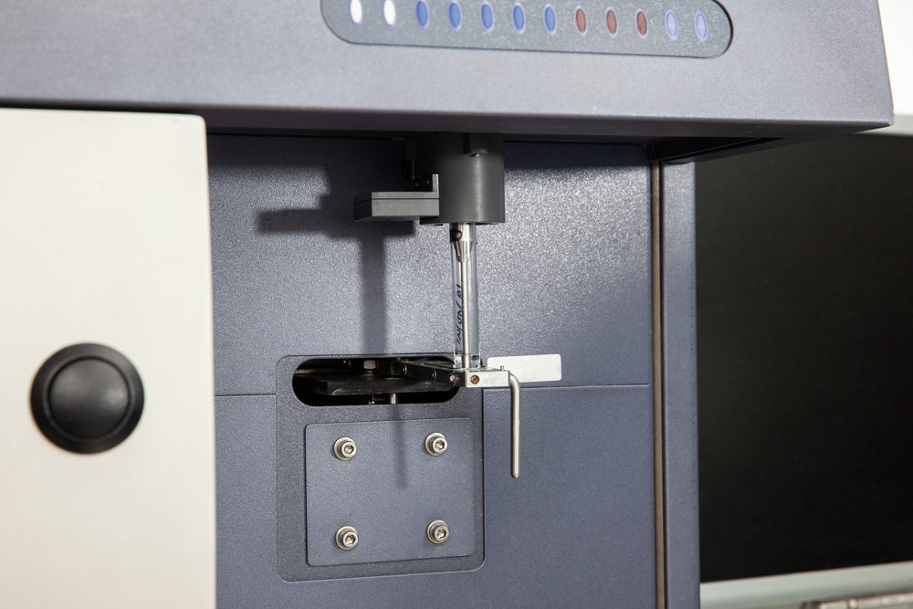
Test tube containing a patient sample loaded on the flow cytometer ready for analysis. Flow cytometer
A popular laser-based technology used by companies like Gentronix, flow cytometry is used to analyse particles or cells. It is a popular method used when it is necessary to analyse the expression of the surface of a cell, as well as the intracellular molecules, defining and characterising different cell types within a heterogeneous population of cells. It is also used to assess the purity of isolated subpopulations and to analyse cell volume and size. Flow cytometry allows for single cells to be analysed in a simultaneous and multi-parameter manner.
Flow cytometry is mainly used to measure the intensity of the fluorescence produced by antibodies detecting fluorescent-labelled proteins or ligands that can bind to specific cell-associated molecules.
The procedure of staining requires a single-cell suspension to be made from cell tissue or culture samples. These cells are then incubated in either microtiter plates or tubes with either fluorochrome-labelled or unlabelled antibodies. This is then analysed using a flow cytometer.
A brief history of the flow cytometer
The first flow cytometer developed for commercial use was introduced in the early 1970s, and in the decades since, it has evolved significantly. By the late 1970s, flow cytometers were using two lasers and had improved capabilities allowing them to measure and quantify cells and adapt for cell sorting. The first spectral flow cytometer was introduced in 2012, using prisms and photomultiplier tubes (PMT) to collect and amplify light.
What are the components of a flow cytometer?
A flow cytometer is made up of three different main components; these are fluidics, optics and electronics.
Fluidics
The responsibility of the fluidics system is for the transportation of cells. This occurs one by one as they travel from the sample tube through the flow cell to a collection tube if this is a case of cell sorters or to the waste.
For illumination to be optimal, the stream used to transport the particles needs to be positioned, so it is at the centre of the laser beam. There should only be a single particle or cell moving through the laser beam at any time. For this to happen, the sample needs to be injected into the stream of sheath fluid found in the flow chamber. On a benchtop cytometer, the flow chamber is called a flow cell. On a stream-in-air cytometer, it is called a nozzle tip. The design of a flow chamber means that the sample core is focused at the centre point of the sheath fluid, where the laser beam interacts with the particles.
Optics
The optics system is necessary for illumination and the collection of light inside the flow cytometer; this includes lenses, lasers, filters and detectors used to convert light to a photocurrent.
Both excitation optics and collection optics form a part of the optical system. The excitation optic comprises both lenses and lasers that are used to focus and shape the laser beam. The collections optic includes a collection lens used to collect the light that the particle-laser beam emits and a system of filters and optical mirrors. These are used to route the wavelength of any collected light to specific optical detectors.
Electronics
The electronic components of a flow cytometer are designed to process and digitise the photocurrent generated from the detector and then save it so it can be analysed.